Only about 65 percent of U.S. women over the age of 40 report having participated in mammography screenings in the past two years. Throughout the EU, 49.2 percent of all women between the ages of 50 and 69 participated in mammography screenings in 2016. However, very large country-specific differences can be observed. The highest participation rates of more than 80 percent were found in Denmark, Finland and Slovenia.
In addition to limited access to mammography, false myths and inadequate knowledge about the procedures could also contribute to the low participation in the screening program. For this reason, practitioners shall provide better and more comprehensive information on the advantages and limitations as well as on the procedures of mammography screening.
What is mammography?
Mammography is an X-ray-based examination of the female breast used to make breast cancer (mammary carcinoma) or its precursors visible. Nowadays, fully digital systems are commonly used; these offer many benefits as compared to the film mammography: immediate availability of the results, improved image quality, and reduced radiation exposure. In the most common screening settings, mammography generates two 2D-images of each breast. However, many radiological praxes, nowadays, offer 3D-images of the breast with techniques of tomosynthesis (a pseudo 3D-imaging technique that uses layered images to create a three-dimensional representation of the breast) or breast CT (which generates real 3D images of the breast without need of breast compression).
When is mammography necessary?
Mammography is used in three situations:
- For early detection of breast cancer, independent of any concrete suspicion (secondary prevention)
- For clarification in cases of a concrete suspicion of breast cancer
- As aftercare for former cancer patients to detect recurrences at an early stage (tertiary prevention)
The American Cancer Society recommends mammography screening for women at average risk of breast cancer with the following frequency:
- between 40 and 44 years: optional once-yearly screening
- between 45 and 54 years: screening once a year
- over 54 years: either annual screening or every two years as long as the woman is in good health and has an expected life expectancy of at least 10 years
Why are informed consent discussions so important?
The mammography screening is the only early detection method that has been proven to reduce the mortality rate from breast cancer. Nevertheless, only about 65 percent of all women in the U.S. regularly take advantage of mammography screening examinations – according to statistics, the participation rate has remained relatively stable between 2008 and 2019. Studies show that benefits are not always accurately assessed. For example, few women are mostly not aware of the possibility of being overdiagnosed.
For this reason, there is an urgent need to educate the population on the benefits and limitations of mammography screening. Studies have shown that information brochures and other similar types of information material have little influence on women’s level of knowledge and willingness to participate. Therefore, a personal consultation with a doctor is the most important way to enable women to make an informed decision.
What should I consider before mammography?
As part of the screening program, patients are entitled to an informed consent discussion with a radiologist. This takes place during a separately scheduled appointment before the mammography to help give women time to make an informed decision and to ask potential questions. Doctors should use this appointment to convey well-founded information and explain the possible advantages and limitations of mammography in an easily understandable manner.
Patients should receive the following information as part of their informed consent discussion:
What is mammography screening?
Mammography screenings are voluntary examinations designed to detect breast cancer at an early stage. Most health insurers cover the cost of the screening – either once a year or every two years – for women over 40. The mammography examination itself is usually performed by X-ray assistants, after which the images are reviewed by trained radiologists.
If abnormalities are detected, the woman is invited for an appointment with an additional examination to confirm or exclude the presence of breast cancer. If the suspicion of cancer is confirmed, which happens only in rare cases, the treating physician will then discuss the further procedure with the patient.
How is mammography carried out?
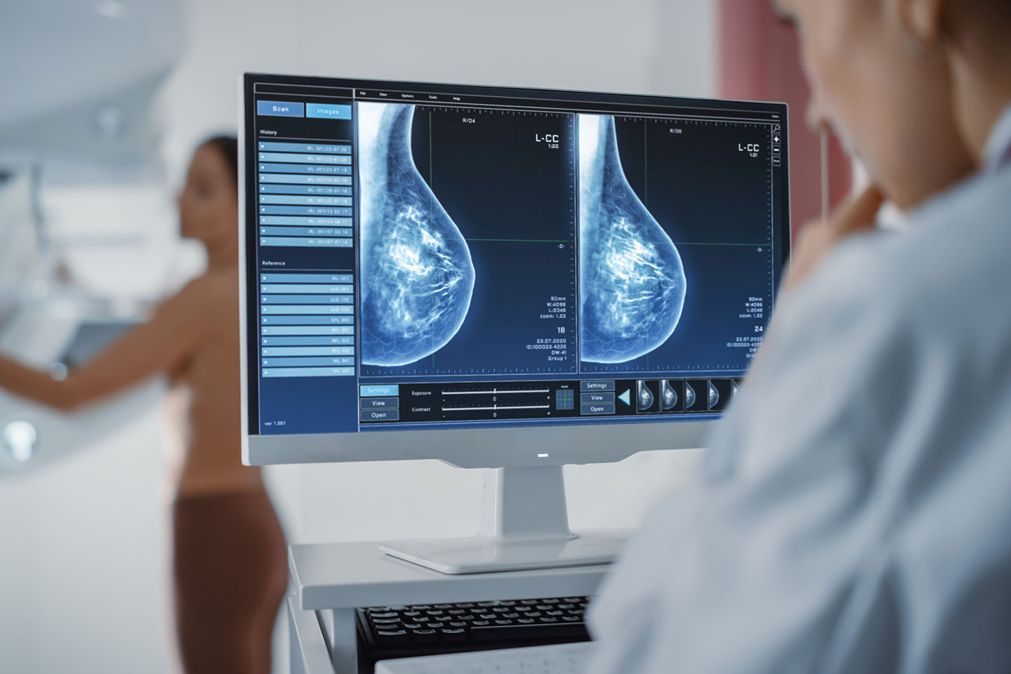
Mammography takes place at an outpatient clinic specializing in the procedure. The examination only takes a few minutes. Each breast is placed between two plates of plexiglass and then pressed as flat as possible. The compression can be for some women somewhat uncomfortable or even painful, but the breast tissue is not damaged. Two X-ray images are then taken: one from the top down and the other laterally from the outside inwards. The X-rays make the internal characteristics of the breast visible, enabling small densities or nodules to be identified.
How should patients prepare for mammography?
Because mammography requires the upper body to be undressed, it is advisable to wear clothing that can be easily removed. On the day of the examination, women should avoid using deodorant and body creams in the armpit and breast areas. These products can leave a film on the skin that can negatively affect image quality.
What are the potential advantages and limitations of mammography in the context of early detection?
Women should be aware that mammography screening has scientifically proven benefits but also certain limitations and risks. The most important advantages of mammography screening are:
- Lower mortality rate: Mammography screening is the only early detection method that is proven to lower the risk of dying from breast cancer. In concrete terms, of every 1000 women who regularly attend screening, approx. 2 to 6 avoid dying from breast cancer.
- Higher sensitivity than palpatory examinations: In contrast to palpatory examinations, mammography can detect exceedingly small tumors, even in the precancerous stage.
- Better therapy outcome: When breast cancer is diagnosed at an early stage, in most cases, the chances of recovery are improved. Also, the course of treatment is less aggressive when the tumor is still small.
However, there are also risks and limitations:
- Low specificity: Mammography can produce false-positive results, meaning the presence of breast cancer is initially suspected but later found not to be the case. In numbers, if 1000 women take part in mammography screening, abnormalities will be identified in 30 of them. Later, in 24 of these 30 women, it will be determined that no breast cancer is present. The seemingly positive findings cause anxiety and uncertainty for patients in the short term.
- Low sensitivity in dense breast: Mammograms of breasts with very dense glandular tissue are difficult to interpret. That’s because both dense breast tissue and abnormal breast changes, such as calcifications and tumors, appear as white areas on the mammogram. As a result, mammography is less sensitive in women with dense breasts—that is, it is more likely to miss cancer. Women with dense breasts may be called back for follow-up testing more often than women with fatty breasts. In many countries in the world, the breast density is indicated in the mammography report. Women with dense breast shall undergo supplemental screening with other imaging methods, such as ultrasound, or MRI.
- Overdiagnosis: In some cases, tumors are found that would never have caused problems, either because they grow very slowly or because women die earlier from other causes. This can result in unnecessary diagnostic procedures and treatments, which may potentially reduce the patient’s quality of life. Furthermore, simply being diagnosed with cancer places an emotional strain on the patient. Of every 1000 women who participate in the screening program, around 9 or 10 of them will receive unnecessary treatment.
- The risk of false-negative findings: In approx. 1 in 10 cases, the presence of cancer will be missed during mammography, despite the high level of care taken. This can give those affected a false sense of security, meaning they pay less attention to symptoms and warning signs.
- Radiation exposure: The radiation waves used during mammography can be cancerous, although the risk is exceptionally low. At the very most, 1 in 1 000 000 women can develop breast cancer because of mammography.( Chijoke et al. Evaluation of mean glandular does and assessment of the risk radiation induced carcinogenesis in women following screening mammography in a low resource setting. Journal of radiation research and applied science. 2018: 11.)
- Breast compression: Compression of the breast spreads out the normal glandular tissue. This makes it easier for radiologists to detect abnormalities. If compression is inadequate, the overlapping tissue may lead to misinterpretation as an abnormality or mass. Novel techniques of breast CT allow the screening of the breast tissue without compression. Women can also discuss alternative screening options with the practitioners.
Patients should also be aware that mammography is a method of early detection (secondary prevention) and is not a method of prevention (primary prevention). This means that mammography cannot prevent the onset of cancer but potentially detect it in the initial stages.
Are there alternative examination techniques to mammography?
Other examination techniques used in the early detection of breast cancer include:
- Regular breast self-examination
- Palpatory examination by a doctor
- Ultrasound examination
- MRI examination (magnet resonance imaging)
These techniques are generally used as complementary examinations alongside mammography. However, none of these alternative techniques have been scientifically proven to reduce the mortality rate associated with breast cancer.
Breast cancer prevention is only successful when coupled with good information
Every woman can and should individually weigh up the advantages and limitations of mammography screening. However, doctors have the task of providing the factual information the patient needs to make an informed decision. Informed consent discussions are also important to ensure that women do not misestimate the benefits of mammography screening. For this reason, the success of breast cancer screening depends not only on the specialist medical knowledge of staff but to a substantial extent on the detailed information provided by the doctor.