It’s a well-known radiology dilemma: Dense breast tissue makes it difficult to detect tumors in X-rays of the female breast. Yet, the risk of carcinoma also rises with breast density. The challenge here is that mammograms become less accurate for women who are more likely to develop breast cancer. So, what are the options for women with dense breast tissue, when it comes to offering them tailored early breast cancer detection? And can these strategies also be implemented for routine screenings?
Definition: What exactly is dense breast tissue?
Female breast tissue is made up of three tissue types: glandular, connective and fatty tissue. Every woman has her own unique proportion of these tissue types. If the relative proportion of glandular and connective tissues happen to be higher, then this is classed as high breast density, sometimes known as a mammographically dense breast. This is because glandular tissue is stronger at absorbing X-rays, while fatty tissue is largely transparent on an X-ray image.
Most tumors are visible via their increased X-ray density, making it easier to detect them via mammograms when the breast density is lower. However, when there is dense breast tissue, this can hide any present tumor tissue, reducing the information gained from the mammogram.
A woman’s breast density depends on several individual factors: genetic predisposition, hormones, body mass index (BMI), alcohol consumption, and even how often the woman has been pregnant. And of course, a major factor is age: as women age, glandular tissue will transform to fatty tissue, reducing the breast density.
Breast density classification according to ACR criteria
The American College of Radiology (ACR) categorizes breast density into four types, in line with BI-RADS assessment guidelines for mammogram results. The newest version (5th ed.), defines these four types as follows:
- ACR A: Almost completely transparent, fatty breast
- ACR B: Loosely distributed parenchyma with increased density
- ACR C: Heterogeneously dense breast with confluent areas of increased density; small tumors may be missed
- ACR D: Almost completely dense breast tissue; reduced mammogram sensitivity
The last two categories, C and D, are usually grouped together as “dense breast tissue”.
Currently, the ACR categories are based on a visual assessment of density via mammogram images. The categories are purely descriptive, in line with BI-RADS guidelines, therefore leaving them to the subjective assessment of the radiologist.
What percentage of women have high breast density?
The ACR cites that mammogram screenings reveal an average of app. 10% of women to have fatty breast tissue (ACR A), 40% have slightly increased (ACR B) or moderately increased breast density (ACR C), while app. 10% have very dense breast tissue (ACR D). This means that about half of all women have a breast density that would cause mammograms to be less effective.
Notably, the research data is based on women that are of typical screening age. Currently, there’s not much data available to confirm whether women under 40 indeed have higher breast density, as mammograms don’t form part of recommended screening methods for this age group.
Dense breast tissue as an independent risk factor for breast cancer
Increased breast density is a risk factor for the development of breast cancer (though moderate). This is because breast tumors usually originate within epithelial structures, such as glandular lobules or milk ducts – hence, denser glandular tissue.
There are different stances on the exact extent to which this risk rises. Older studies cite that dense breast tissue increases the risk of breast cancer by a factor of 4 to 6. A more recent meta-analysis concludes that women with very high breast density (ACR D) have an increased cancer risk by a factor of 2, in comparison to women with slightly increased breast density (ACR B). The difference in these estimates may be down to changing ACR criteria over time, as well as the switch from using film to digital mammogram systems.
Yet even if the risk is lower than previously thought: Without a doubt, women with dense breast tissue are more likely to develop breast cancer.
Breast cancer diagnostics with dense breast tissue: Which methods are useful?
Aside from being a risk factor, dense breast tissue also makes it more difficult to detect breast cancer in the first place. Mammograms, which are usually the first port of call for screening, become less accurate with higher breast density. With the many imaging techniques available, each has their own strengths and weaknesses when it comes to assessing dense breast tissue.
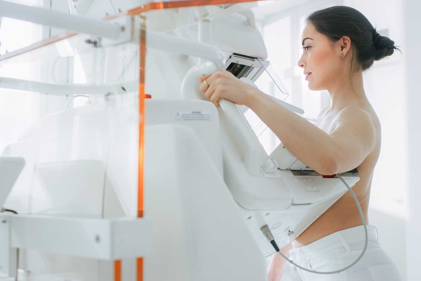
Mammograms
Since dense breast tissue can mask tumors, mammograms become less sensitive with more dense breast tissue. This reduces specificity since pathological findings can be misread due to layering and superimposition in the images.
Despite these faults, mammograms are still crucial parts of basic breast exams – not least because they provide an initial way to assess breast density. Mammograms are also good for identifying microcalcifications as an indication of tumors, even with higher breast density.
Tomosynthesis
Tomosynthesis is a 3D imaging technique using image slices, where the breast tissue is depicted free of overlay. It’s a method better suited to identifying small focal findings and architectural defects over conventional mammograms. A recent TOSYMA study investigated how this can help women with dense breast tissue, giving promising results. Participants with very dense breast tissue benefited from a 250% higher tumor detection rate than with conventional digital mammograms.
Magnetic Resonance Imaging (MRI)
While mammography and tomosynthesis are both X-ray-based methods, detecting differences in density, MRIs reveal a different view: Tumor tissue is detected by its absorbency in contrast agents, due to the tissue’s altered metabolic activity. Thus, contrast-enhanced MRIs are significantly less affected by dense breast tissue than X-ray-based methods.
A Dutch DENSE study shows that additional breast MRIs can significantly reduce the rate of interval tumors in women with dense breast tissue. However, a possible disadvantage of MRIs is the lower specificity, which can lead to a higher rate of false-positive findings.
Ultrasound
Ultrasound exams are already a routine method for clearing up inconclusive findings from mammograms. Recent studies indicate that additional ultrasound testing can detect more tumors in women with dense breast tissue – but this also comes with an increased risk of false positives. A possible advantage of ultrasounds over MRIs – particularly for early detection – is that it takes less time and is less expensive. However, the reliability of an ultrasound is heavily dependent on the experience of the medical staff carrying it out.
Mammogram screenings and dense breast tissue: the challenges
Standardized mammogram screenings, common in the US and many European countries, have proven to reduce breast cancer mortality. Nevertheless, these screening programs still have their weaknesses – especially for women with dense breast tissue.
To optimize breast cancer screening for this patient group as well, there are three main challenges to overcome:
1. Standardized dense breast tissue classification
The ACR may be an internationally recognized classification form of breast density, but this is based on a subjective description of radiologists assessing a mammogram. According to the current 5th ed. of BIRADS, this form of classification is purely descriptive, as the percentages that were once included in the 4th ed. have been abandoned. This means there is still no objective criteria for standardized breast density classification.
The challenge here is that recommendations for further diagnostic procedures depend heavily on subjective assessments by physicians.
2. Mammogram sensitivity to dense breast tissue.
Mammograms have become the primary form of exam for routine breast cancer screenings due to their wide availability, ease of use, and low cost. However, mammogram sensitivity falls with increasing breast density: it plummets from 86-89% in women with fatty breasts (ACR A) to app. 62-68% for very dense breast tissue (ACR D). As a result, women with more highly dense breast tissue suffer from more interval carcinomas – tumors that arise between two screening appointments, or which may have been missed at the first appointment.
3. Standard procedures for dense breast tissue
There is currently no international consensus on how to deal with higher breast density during routine screenings. In the US, the FDA ruled in March 2023 that women must be told about their breast density in a diagnostic setting. However, the FDA doesn’t make any concrete recommendations for further diagnostic procedures.
In its latest recommendations, the European Society of Breast Imaging (EUSOBI) advocates for educating women about their breast density. This involves the offer of additional MRIs at two- to four-year intervals if they have dense breast tissue.
However, this EUSOBI guideline isn’t currently implemented in most European countries. In Germany, for example, during routine mammogram screenings, breast density is neither documented nor communicated to the women. In Switzerland, where screening programs are organized on a regional basis, the procedure is also inconsistent. In general, women with dense breast tissue are told to have additional ultrasound exams.
Optimized breast cancer screening through software-based solutions
It’s clear that the big picture challenge is to standardize – both the classification of breast density as well as the further diagnostic procedures. But how can nations achieve this as part of routine screenings, giving limited financial resources and chronic understaffing?
A key solution could be software-based. The integration of smart algorithms has already made it possible to determine the precise breast density, automatically and on a quantitative basis. Thus, these algorithms help build a standardized risk assessment.
In this way, physicians can make tailored decisions based on objective facts as to whether their patient should receive an additional ultrasound or an MRI. In the future, this software may even be able to provide recommendations for further diagnostic procedures.
Outlook: Individually tailored screening strategies
In order for breast cancer screening programs to benefit individual patients even more, it’s crucial to correctly assess and consider their individual cancer risk. Here, breast density is a key factor. Women with dense breast tissue could benefit greatly from tailored screening strategies – and for this, the right technological solutions need to become part of routine screenings.